Table of Contents
As we already know cell is a very important topic not only for class 9th but also for further classes. It is the topic of the most complex and a little confusing chapter called the Fundamental Unit of Life: Cell from the Biology section of class 9th. So we have explained the animal cell briefly in our article. A cell is the smallest unit of life that can carry out various processes that are necessary for an organism to survive.
The cell is the fundamental, organizational, and structural unit of life. Cells can vary in size, shape, and function, but they typically contain genetic material (DNA or RNA) and are surrounded by cell membranes. All the organisms present on the earth are divided into two types: Prokaryotes and Eukaryotes. Eukaryotes are the ones who have advanced and complete cells. The eukaryotes also contain two types of cells: The plant cell and the Animal cell. In this article, we will be studying Animal cells briefly.
Animal Cell Definition
An animal cell is a remarkable building block of life, forming the basis of every organism. These cells are intricate structures, each with a complex system of organelles that collaborate to ensure the cell’s survival and functionality. The animal cell does not have a rigid cell wall which makes it different from the plant cell. Instead, they have a plasma membrane that regulates the movement of substances in and out of the cell and maintains the internal balance.
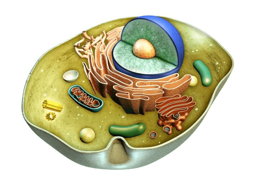
Animal cells are intricate marvels of Biology, each organelle contributing to the cell’s survival and specialized functions. These animal cells are mostly smaller in size, except in the protozoan Euglena no animal cell consists of plastids. The animal cell also lacks chloroplast which is present in the plant cells, and helps the plant cell prepare their own food.
Animal Cell Diagram
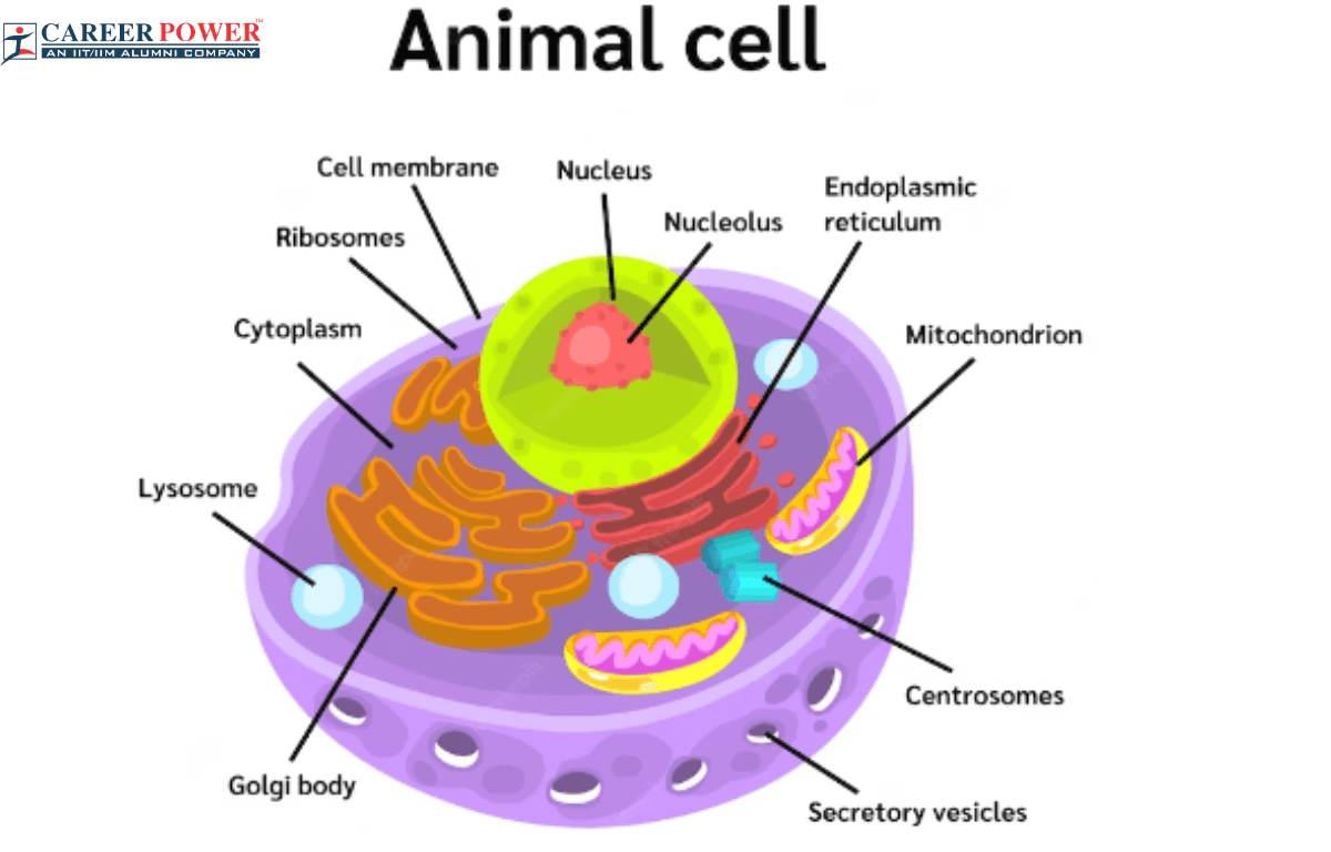
However, this animal cell diagram does not show any one particular type of cell organelle. But gives an overview of all the primary organelles and internal structure of the animal cell. The animal cell diagram depicts the various parts and structures within a cell, including the Nucleus, Cytoplasm, Cell Membrane or Plasma Membrane, Mitochondria, Endoplasmic Reticulum, Golgi apparatus, and many more.
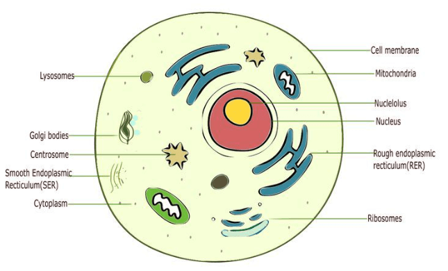
Structure of an Animal Cell
An animal cell is a complex structure with several key components. The cell membrane forms the outer boundary, regulating the passage of substances. Inside, the cytoplasm, a gel-like substance, hosts various organelles. The nucleus, the cell’s control center, contains genetic material (DNA) and regulates cell activities. Mitochondria generate energy, while the endoplasmic reticulum aids in protein and lipid synthesis. The Golgi apparatus processes and packages molecules, and lysosomes break down waste. The cytoskeleton provides structural support, and centrioles aid in cell division. These components collaborate to ensure the cell’s survival and functioning.
Functions of Animal Cell
There are various cell organelles that are present in a cell, and each cell organelle in the animal cell performs many important roles in the proper functioning of the animal body. Some of them are mentioned below:
- With the help of cellular respiration. the mitochondria play a role in the generation of energy Respiration.
- The ribosomes help in the synthesis of proteins using instructions from DNA.
- The nucleus houses DNA, storing genetic information for cell activities and reproduction.
- The plasma membrane controls the movement of substances in and out of the cell, facilitating communication with the environment.
- The enzymes are present in the lysosomes that help break down waste materials and cellular debris.
- The cytoskeleton provides structural support and helps in maintaining the cell’s shape.
- Different cell types perform specialized tasks in tissues and organs, such as nerve cells transmitting signals or muscle cells contracting.
- The animal cell divides through processes like mitosis and meiosis which helps in growth and Reproduction.
- Vacuoles store water, nutrients, and ions which helps in regulating cellular water balance.
- Certain cells of the immune system, like white blood cells, help defend against pathogens and infections.
Parts of Animal Cell
An animal cell consists of many different regions and each region has its own role to play. Based on their shapes, size, and activities all the cells have the following major functional parts: The plasma membrane, nucleus, cytoplasm, and cell organelles. Here we have discussed all of these parts briefly in a tabulated form:
| Different Parts of an Animal Cell | |
| Name of the parts | Description |
| Plasma Membrane | A thin, flexible barrier that surrounds and protects the cell. It controls the movement of substances in and out of the cell. |
| Nucleus | The nucleus is the cell’s control center, which holds the DNA and all the directing activities. It is enclosed by a double membrane and contains the nucleolus and chromatin. |
| Cytoplasm | A gel-like substance within an animal cell, where various cellular activities take place. It holds all the cell organelles and provides a medium for chemical reactions. |
| Cell Organelles | Each cell organelle performs a specific role within the animal cell. There are several cell organelles in an animal cell. some of the key organelles are mitochondria, ribosomes, lysosomes, cytoskeleton, Golgi apparatus, and many more. |
1. Plasma membrane
The plasma membrane also known as the cell membrane is a vital component in an animal cell. The plasma is the outer boundary, which encloses the cell, separating its internal structures from the external environment. The plasma membrane is constituted of a lipid bilayer embedded with various proteins, which play crucial roles in maintaining the cell structure and controlling the movement of substances in and out of the cell.
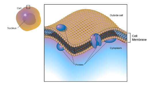
Functions of a Plasma membrane
- The plasma membrane helps to interact with the neighboring cells for the transmission of signals.
- The plasma membrane controls the entry and exit of the molecules.
- It helps in the regulation of the pathways so that the substances can pass through easily.
- The plasma membrane also provides structural support to the animal cell.
2. Nucleus
The nucleus is the membrane-bound cell organelle that contains all the cell’s genetic material like DNA, which contains instructions for making proteins and controlling various cellular processes. The nucleus plays a crucial role in the cell’s functioning. A nucleus is surrounded by a double membrane called a nuclear envelope, which has nuclear pores allowing communication between the nucleus and the rest of the cell. Inside the nucleus, there is a nucleolus that helps in the synthesis of ribosomes and chromatin, which contains DNA and protein and undergoes changes to regulate gene expression.
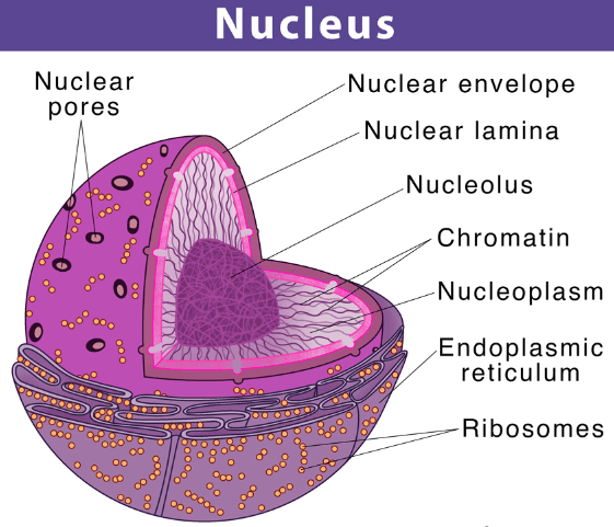
Functions of Nucleus
- The nucleus helps in the regulation of the cell cycle.
- If the nucleus is removed from the cell, the protoplasm ultimately dries up and dies.
- It also controls the transmission of heredity traits from the parents to the offspring.
- All the metabolic activities of the cell are controlled by the nucleus.
3. Cytoplasm
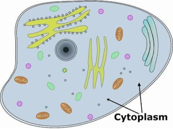
Cytoplasm is like the jelly inside a jelly-filled donut for a cell. It is a gel-like substance that fills up the space between the cell membrane (the donut’s outer layer) and the nucleus (the donut’s center). Cytoplasm is where many important activities happen in a cell, like breaking down nutrients for energy, making proteins, and storing materials. It’s filled with tiny structures called organelles, like mitochondria (powerhouses of the cell) and ribosomes (protein factories). Cytoplasm also helps maintain the cell’s shape and provides support to organelles.
Functions of Cytoplasm
- Living cytoplasm is always in a state of movement.
- The cytoplasm stores various chemicals such as amino acids, glucose, vitamins, ions, etc.
- The cytoplasm is the site of certain metabolic pathways, such as glycolysis.
4. Cell Organelles
There are a lot of cell organelles found in a single animal cell. Ans each organelle has its own important role. The cell organelles are specialized structures within the cell that perform distinct functions. All the cell organelles work together to ensure a cell’s survival and functioning. The nucleus houses the DNA and controls the cell’s activities, while mitochondria generate energy. The ER synthesizes proteins and lipids, which are processed and packaged by the Golgi apparatus. The lysosomes break down the waste and vacuoles store materials. The cell membrane regulates the passage of the substances. The different cell organelles that are found in the animal cell are:
- Endoplasmic Reticulum
- Mitochondria
- Lysosomes
- Vacuoles
- Ribosomes
- Golgi Apparatus
- Centrioles
- Cytoskeleton
- Peroxisome
Functions of Cell Organelles
We have discussed the functions of a few cell organelles here. These organelles work together to ensure the power functioning and survival of the cell.
- Nucleus: The nucleus contains genetic information (DNA) and controls the cell’s activities, including growth and reproduction.
- Mitochondria: Mitochondria are the powerhouse of the cell, producing energy (in the form of ATP) through cellular respiration.
- Endoplasmic Reticulum: The endoplasmic reticulum ER is involved in the synthesis and transport of proteins and lipids.
- Golgi apparatus: The Golgi apparatus modifies, sorts, and packages proteins and lipids for storage or transport out of the cell.
- Lysosomes: Lysosomes are responsible for breaking down waste materials and cellular debris, and recycling components of the cell.
- Cytoplasm: The cytoplasm is a gel-like substance that fills the cell and provides a medium for the organelles to be suspended in. It also facilitates cellular processes.
- Cell Membrane: The cell membrane (plasma membrane) regulates what enters and exits the cell, maintaining the cell’s internal environment.
- Ribosomes: Ribosomes are the sites of protein synthesis in the cell, where amino acids are assembled into proteins based on the instructions from RNA.
- Vacuoles: In plant cells, vacuoles store nutrients and waste products, helping maintain turgor pressure. In animal cells, they are smaller and primarily store nutrients and waste.
1. Endoplasmic Reticulum
The endoplasmic reticulum is a membranous and complex cell organelle in animal cells involved in various functions. It plays many important functions inside the cell. The endoplasmic reticulum consists of a network of membranous tubules and flattened sacs called Cisternae. The Endoplasmic Reticulum can be classified into two main types :
- Rough Endoplasmic Reticulum
- Smooth Endoplasmic Reticulum
For more details, you can also go through our article on the Difference between Rough endoplasmic and Smooth endoplasmic reticulum.
Rough Endoplasmic Reticulum
- The rough endoplasmic reticulum contains ribosomes attached to its surface, which give the ER a “rough” appearance under a microscope.
- The ribosomes on the Rough ER synthesize proteins that are either secreted from the cell, incorporated into the cell membrane, or sent to lysosomes.
- As proteins are synthesized, they enter the lumen of the RER, where they undergo folding, modification, and quality control.
Smooth Endoplasmic Reticulum
- The smooth endoplasmic reticulum lacks ribosomes on its surface, which provides it with a “smooth” appearance under a microscope.
- The smooth endoplasmic reticulum functions in various metabolic processes, which include lipid metabolism, and detoxification of drugs and toxins.
- The smooth ER stores calcium ions that are critical for muscle contraction, cell signaling, and other processes.
- It is also involved in the metabolism of carbohydrates and steroids.
2. Mitochondria
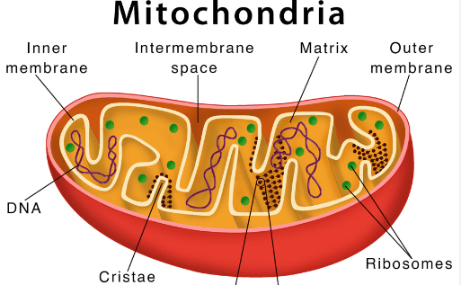
The mitochondria are also known as the “Power House of the Cell”. The mitochondria help in the production of energy in the form of ATP through the process known as cellular respiration. This energy is vital for the proper functioning of the cell. Accept all other cell organelles the mitochondria have their own DNA and ribosomes. The mitochondria play a crucial role in energy production, metabolism, and cell survival.
3. Golgi Apparatus
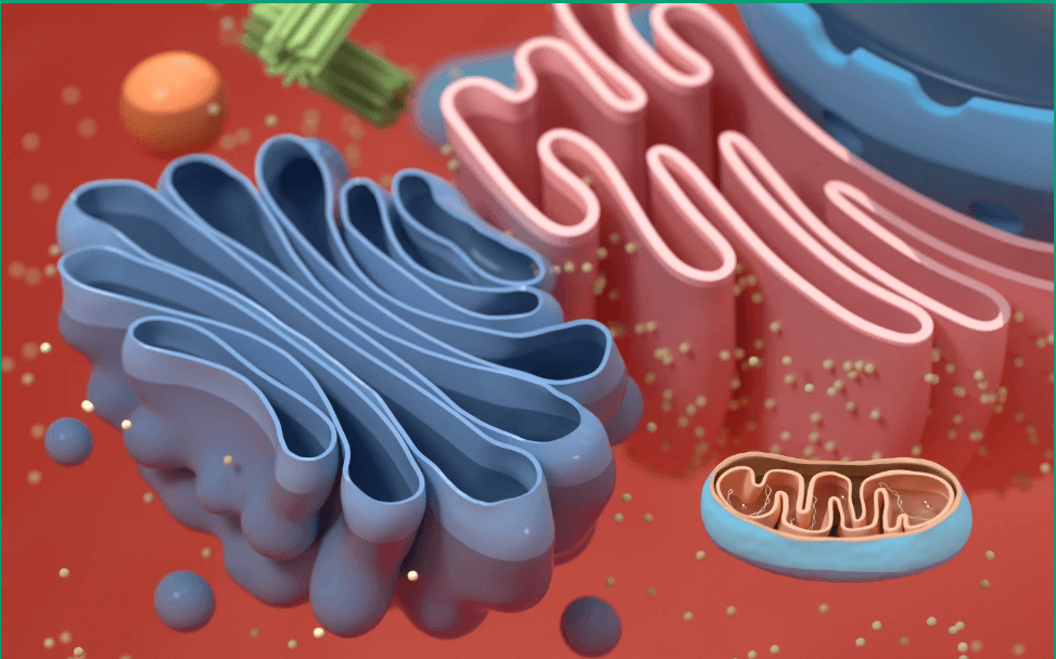
The Golgi apparatus is generally known as the Golgi complex, an organelle found in both plant and animal cells. The Golgi apparatus helps in the modification and packaging of proteins and lipids for transport within and outside the cell. The Golgi apparatus acts like a post office, which helps in the preparation of molecules for delivering them to their proper destination. The Golgi apparatus consists of a series of flattened, membrane-bound sacs called cisternae.
4. Lysosomes
The lysosomes are membrane-bound cell organelles that are found in the animal cell. The lysosomes are sac-like organelles that contain enzymes that break down waste material and cellular contaminants. Lysosomes are often referred to as the “garbage disposal” or recycling center of the cell. Lysosomes play a very important role in the removal of damaged organelles, recycling cellular components, and breaking down foreign substances that may enter the cell. The lysosomes are also known as the suicidal bags of the cell. When a cell gets damaged the lysosomes release digestive enzymes. These enzymes then eat up their own cell.
5. Vacuoles
In animal cells, vacuoles are smaller and less prominent compared to the plant cell. Animal cell vacuoles are typically smaller membrane-bound structures. These vacuoles are storage sacs that can contain water, nutrients, and other waste products. They play a very crucial role in various cellular processes such as sorting waste material, maintaining cell volume, and assisting in intracellular digestion.
6. Centrioles
The centrioles are cylindrical structures found in animal cells. They are typically found near the nucleus of the cell. The centrioles are involved in cell division and are particularly important during the process of cell replication. They are responsible for the formation of spindle fibers that help to separate chromosomes during mitosis and meiosis. Centrioles are composed of microtubules and are often organized in pairs called centromeres.
7. Peroxisomes
The peroxisomes are the membrane-bound organelles that are found in animal cells. The peroxisomes are involved in breaking down fatty acids and detoxifying harmful substances. Additionally, the peroxisomes play a crucial role in various metabolic processes, such as the production and breakdown of hydrogen peroxide, which is a potentially harmful byproduct of certain metabolic reactions. The peroxisomes help in the maintenance of the cell’s overall health and functions.
8. Ribosomes
The ribosomes are cellular structures that are found in both plant and animal cells. These ribosomes are tiny structures that are responsible for protein synthesis, translating genetic information from RNA into proteins. The ribosomes consisted of two subunits: a larger one and a smaller one. These subunits are composed of ribosomal RNA (rRNA) molecules and proteins. The ribosomes are found free in the cytoplasm or attached to the endoplasmic reticulum, it depends on the type of protein they synthesize.
9. Cytoskeleton
The cytoskeleton of an animal cell is a dynamic network of protein filaments and tubules that provides structural support, helps in the maintenance of the cell’s shape, and enables various cellular processes. The cytoskeleton is a set of three main components that are: Microfilaments, Intermediate filaments, and Microtubules.
Microfilaments
The microfilaments are thin, thread-like structures made of actin proteins. They help with cell movement and support cell shape. The microfilaments are involved in processes like muscle contraction and cell division.
Intermediate Filament
The intermediate filaments are slightly thicker filaments made up of various proteins depending on the cell type. The intermediate filament provides mechanical strength to the cell and helps anchor organelles in place.
Microtubules
The microtubules are hollow tubes made up of tubulin proteins. They are important for maintaining the cell shape, forming tracks for intracellular transport, and segregating chromosomes during cell division.


 Parts of a Tree, Names and Their Functio...
 CBSE Class 12 Biology Viva Questions Wit...
 Binary Fission, Step by Step Process in ...


